Journal of Bioscience and Bioengineering Vol. 102, No. 2 (2006)
Vol. 102, August 2006
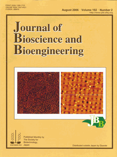
Tapping mode atomic force microscopy (AFM) topographic images of L3-liposome.
The AFM images of electron-beam-developed areas in the scales of 30 x 30 μm2 (left) and 5 x 5 μm2 (right). L3-liposome was prepared on the patterned substrate by electron-beam lithography technique. This method is greatly anticipated for biosensor application.
Related article: Jung, H. S., Kim, J. M., Park, J. W., Lee, S. E., Lee, H. Y., Kuboi, R., and Kawai, T., “Atomic force microscopy observation of highly arrayed phospholipid bilayer vesicle on a gold surface“, J. Biosci. Bioeng., vol. 102, 28-33 (2006).
⇒JBBアーカイブ:Vol.107 (2009) ~最新号
⇒JBBアーカイブ:Vol. 93(2002)~Vol. 106(2008)


.gif)