Journal of Bioscience and Bioengineering Vol. 97, No. 6 (2004)
Last updated: 2022.04.18
Vol. 97, June 2004
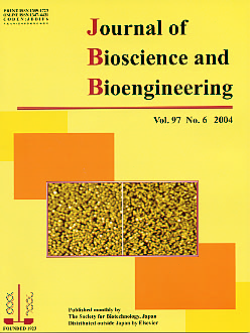
Typical atomic force microscopy images of the streptavidin layers adsorbed on a mica surface (left panel) and the pretreated gold surface (right panel) in the originally scanned 1 x 1 μm2 areas.
Related article: Kim, J., Yamasaki, R., Park, J., Jung, H., Lee, H., and Kawai, T., "Highly dense protein layers confirmed by atomic force microscopy and quartz crystal microbalance", J. Biosci. Bioeng., vol. 97, 138-140 (2004).
⇒JBB Archive Top
⇒JBB Archive: Vol. 93 (2002)–Vol. 106 (2008)
.jpg)